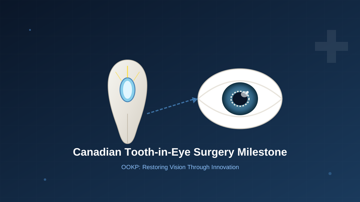
Canadian Medical Milestone: First Tooth-in-Eye Patients Regain Sight After Groundbreaking Surgery
In a remarkable achievement for Canadian medicine, three completely blind patients have regained their sight through an extraordinary surgical procedure that uses their own teeth as the foundation for restoring vision. The operations, performed at Mount Saint Joseph Hospital in Vancouver, represent the first time this rare technique has been successfully completed in Canada.
The procedure, known as Osteo-Odonto-Keratoprosthesis (OOKP) or more commonly "tooth-in-eye surgery," was pioneered by Italian ophthalmologist Benedetto Strampelli in the 1960s. Despite its six-decade history, it remains one of the rarest and most complex eye surgeries in the world, with only a handful of centers globally offering this life-changing intervention.
The Patients and Their Journey
Among the three pioneering Canadian patients is Gail Lane, a 75-year-old Victoria resident who lost her sight at age 64. Her blindness resulted from Stevens-Johnson Syndrome, a severe autoimmune disorder triggered by anti-seizure medication she had been prescribed. The condition extensively scarred her corneas, plunging her into darkness after a lifetime of normal vision.
Brent Chapman, a 34-year-old from North Vancouver, represents another success story. Having lost his sight as a teenager, Chapman underwent the same procedure with remarkable initial results. A third patient, who chose not to be publicly identified, also benefited from this groundbreaking surgery.
How the Procedure Works
The OOKP procedure consists of two separate surgeries spaced approximately three months apart, each requiring extraordinary surgical precision and interdisciplinary collaboration.
In the first surgery, the patient's own canine tooth—specifically the eye tooth—is carefully extracted. A tiny optical cylinder with an artificial lens is inserted into a precisely drilled hole in the center of the tooth. This tooth-lens complex is then implanted into a pocket created in the patient's cheek. Over the following three months, living tissue grows around and into the tooth, creating a biological housing that will eventually support the optical implant.
The biological integration is crucial. The tooth's hard enamel provides an inert, stable platform that the body will not reject, while the surrounding tissue creates a vascularized biological environment that can sustain the implant. Attempting to place the tooth directly into the eye without this preparatory step would be impossible due to tissue rejection and inadequate support.
In the second surgery, performed after the three-month healing period, the tooth-lens complex is carefully removed from the cheek and implanted into the patient's eye. This five-hour procedure requires the surgeon to create a precise opening in the damaged cornea, remove the opaque tissue, and secure the tooth-lens assembly in place. The surrounding tissue is then carefully reconstructed around the implant.
Recovery and Results
Each patient's recovery followed a unique trajectory, demonstrating the individualized nature of healing after such complex surgery.
Gail Lane's vision returned gradually over several weeks. Initially, she only perceived increased light sensitivity in her left eye. Slowly, she began distinguishing larger objects, though they appeared fuzzy and indistinct. Then in mid-July, her vision dramatically improved—she could see definition, type, and size of objects and people.
The first object she clearly saw was her partner's black Labrador guide dog, Piper. She could see the dog's tail wagging, and gradually the rest of the animal came into view. Shortly after, she saw her partner Phil for the first time in over a decade. With prescription glasses, Lane's vision was measured at 20/50—a remarkable improvement from complete blindness.
Brent Chapman experienced even more dramatic immediate results. About a week after his final surgery, he achieved 20/30 vision with corrective lenses. However, as sometimes happens during healing, the lens-tooth complex became slightly misaligned, affecting the visual signals reaching his optic nerve. A minor adjustment surgery was planned to reposition the lens, with excellent expected outcomes.
The third patient experienced perhaps the most remarkable immediate recovery—able to see the day after the final surgery and capable of reading documents.
The Surgical Team and Support
The procedures were led by Dr. Greg Moloney, a surgeon and ophthalmologist who brought the OOKP technique to Canada after performing eight similar operations in his native Australia. Dr. Moloney was recruited to Mount Saint Joseph Hospital four years ago specifically to establish this capability for Canadian patients.
The surgical success relied on extensive teamwork. "I am so proud of the surgical team, nursing and hospital staff that made these surgeries possible," Dr. Moloney stated. The operations required coordination between ophthalmologists, oral surgeons, anesthesiologists, and specialized nursing staff.
Critical financial support came from St. Paul's Foundation, which raised CAD 430,000 to fund the OOKP clinic for three years. This funding covered start-up costs, specialized training, equipment acquisition, and yearly operating expenses necessary to establish this rare capability.
Impact on Patients' Lives
The return of vision represents more than improved eyesight—it fundamentally transforms daily existence and restores independence.
Gail Lane describes the practical changes with characteristic candor. She no longer needs to call volunteer services like the Be My Eyes app to verify whether her socks match or whether her outfit is coordinated. Simple pleasures like eating at a restaurant without embarrassment have returned. As a blind person, she would often lift a fork of salad to her mouth only to discover nothing was there—salad is so light that without vision, she couldn't feel it on the fork.
Lane is looking forward to walking a few blocks to the store without depending on someone else's assistance. She is eager to return to playing mahjong with friends and reading books rather than relying solely on audio versions.
"It's like a miracle to me," Lane says, becoming emotional when recalling her decade-long journey. "It was a long, long road and I couldn't have done it without the medical team and all my family and very good friends. It's a special and rare procedure, but it's a wonderful thing."
Who Qualifies for OOKP Surgery
Osteo-Odonto-Keratoprosthesis is not a first-line treatment for blindness. It is specifically designed for patients with severe bilateral corneal blindness who have failed all other conventional treatments, including corneal transplants.
Ideal candidates include patients with:
- Stevens-Johnson syndrome (as in Gail Lane's case)
- Chemical or thermal burns to the eyes
- Multiple failed corneal transplants
- End-stage ocular surface disease
- Certain autoimmune conditions affecting the cornea
Critical requirements include having at least one healthy tooth suitable for extraction and a functional retina and optic nerve capable of transmitting visual signals. The procedure cannot help patients whose blindness originates from retinal or optic nerve damage.
Global Context and Availability
Before these Canadian surgeries, patients seeking OOKP had to travel to specialized centers in the United Kingdom, Italy, India, Australia, or Singapore. The establishment of this capability at Mount Saint Joseph Hospital makes Canada one of fewer than twenty countries worldwide offering this procedure.
The rarity of OOKP reflects its complexity and the specialized training required. The surgery demands expertise in both ophthalmic and dental surgery, as well as extensive experience with the specific techniques developed over decades of refinement since Strampelli's original work.
Looking Forward
Dr. Moloney emphasizes that these initial successes represent the beginning, not the end, of the journey. "Our first big goal was to finish the surgeries without complications and have the back of the (patients') eye healthy enough for them all to see again. We did that. The next goal is to make sure the prosthetics (teeth) are viable in the long term."
OOKP prosthetics require lifelong monitoring and care. The biological integration between the tooth and surrounding tissue must be maintained, and the optical cylinder must remain properly positioned. Patients need regular follow-up appointments and must follow strict protocols to protect their new vision.
Nevertheless, for patients who have exhausted all other options, OOKP represents a genuine miracle—transforming years of darkness into the possibility of seeing loved ones, navigating independently, and reclaiming the simple joys of daily life.
Sources and Further Reading
- The Daily Scan - Providence Health Care
- Providence Health Care - Mount Saint Joseph Hospital
- Strampelli B. (1963). Osteo-odontokeratoprosthesis. Ann Ottalmol Clin Ocul.
